- Overview
- Features
- Specification
Basic Info.
Model NO.
XrESWL-XTS
Penetration Depth
130mm
Voltage Range
12-20kv
Power Supply
220V, 50kHz
Energy Level
0-20
Frequency
45-90/Minute
Certificate
ISO, Ce
Warranty Period
2 Years
After-Sales Service
Whole Using Lifetime
Transport Package
Wooden Case
Specification
2000mmx665mmx700mm
Trademark
XINDRAY MEDICAL
Origin
China
Production Capacity
10000
Product Description
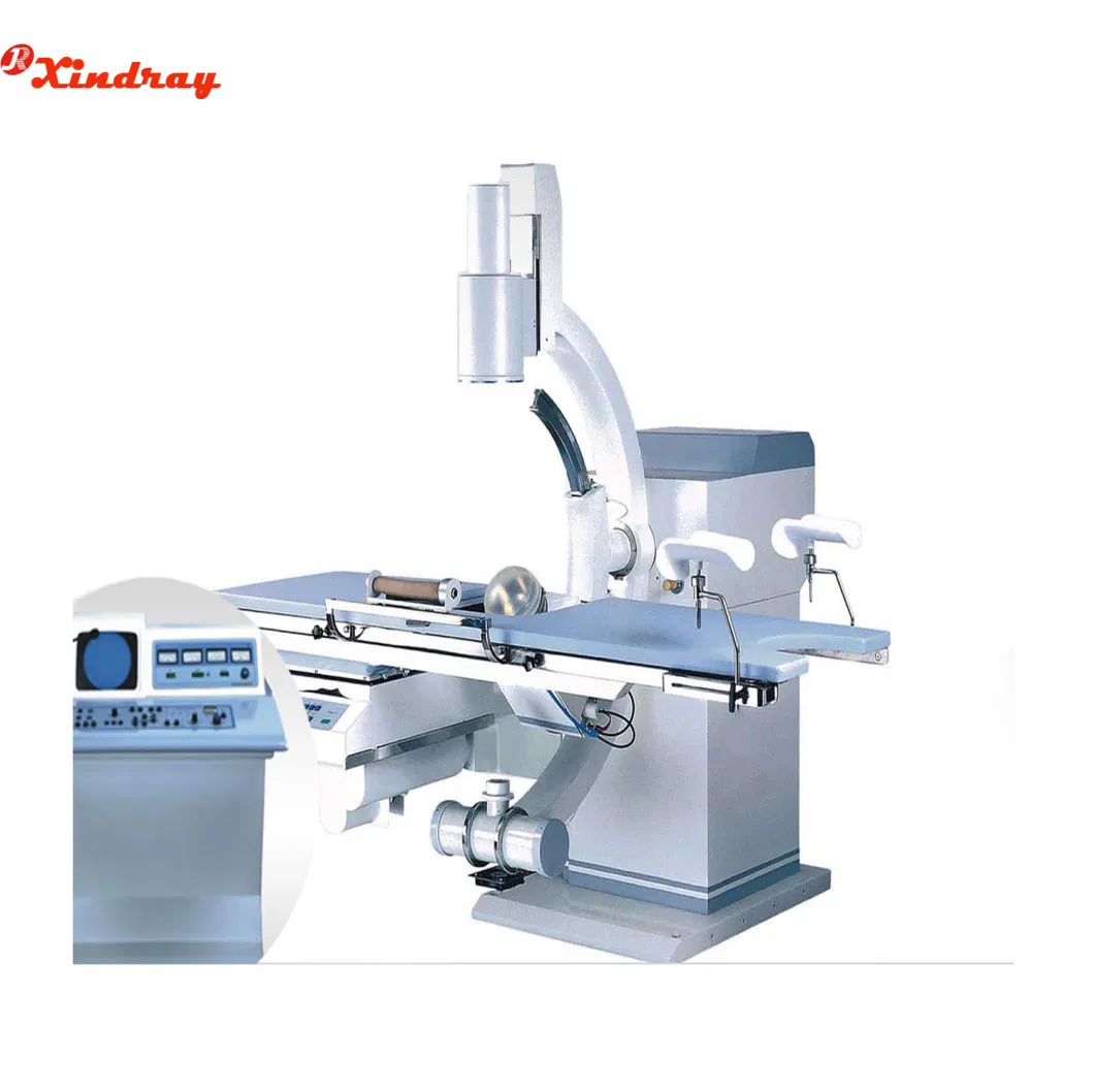
Hospital Medical Equipment X-ray Position Urology ESWL Machine

Application scope Urinary system calculus
Environmental condition
1,Ambient temperature 15~35ºC
2,Relative humidity 45~85%
(1) Main room area ≥4m×4m×2.5m
(2) Operate room area ≥2m×4m.
(3) Main room request equip with X-ray protection function
Electromagnetic shock wave generator
1,High-voltage discharge range: 5~11KV
2,Energy level: 5~24J
3,The second focus shock wave pressure field parameters:
(1) Plus front ≤0.5us
(2) Plus width ≤1us
(3) Focal zone Radial±7.5mm,Axial±12.5mm
(4) Shrinking pressure: 20-50MPa
(5) Penetration ≥130mm
4,Shock wave generator move spherical centered on focus area.
5,Shock wave generator adopted EM shock wave source
Locate system
1, X-ray locate system(C -arm single X-ray two way locate)
(1) X-ray tube voltage 50~110KV;
(2)X-ray tube current ≤5mA;
(3)X-ray image definition ≥14LP/ mm;
(4) High resolution image intensifier and high definition CCD camera.
2,B-ultrasound locate system
(1) Probe axis line
Probe do linear motion around focal point caused inaccuracy ≤±2mm
Probe do circus movement around focal point caused inaccuracy ≤±2mm
(2) Inaccuracy between probe surface and the second focal point <2mm
Certificate:

Package:

Shipping:

Our Service:


Features:
Usage:
Orthopaedics:osteopathy, diaplasis, nailing
Surgery: removing foreign body, cardiac catheter, implanting pace maker, interventional therapy, partial radiography, local photography, and other work.
Orthopaedics:osteopathy, diaplasis, nailing
Surgery: removing foreign body, cardiac catheter, implanting pace maker, interventional therapy, partial radiography, local photography, and other work.
- With a compact appearance, and easy to operate
- Unique base electric auxiliary support arm design, it's more security for using.
- A unique hand-held controller design, convenient to operate
- With a high-quality knockdown X-ray generator to reduce radiation
- With the Perspective KV,MA automatically track fluoroscopy to make the image brightness and clearness optimum
- Toshiba image intensifier, the quality is stable and reliable, a good image clarity
- Clinical Video system with high performance,8 images storage volume, and three 14inchhigh-resolution monitors
- Installation of dense grain grids, to further enhance image sharpness
Specifications:
| High-voltage discharge range: | 5~11KV |
| Energy level: | 5~24J |
| Shrinking pressure: | 20-50MPa |
| Penetration | ≥130mm |
| X-ray tube voltage | 50~110KV |
| X-ray tube current | ≤5mA |
| X-ray image definition | ≥14LP/ mm |
| Operate room area | ≥2m×4m |
Application scope
Environmental condition
1,Ambient temperature 15~35ºC
2,Relative humidity 45~85%
3.Atmospheric pressure 86~106kPa
4.Power requirements AC 3N~220V±10% 50±1Hz, PW≤5kW
5.Water requirements: demineralized water
6.Space requirements(1) Main room area ≥4m×4m×2.5m
(2) Operate room area ≥2m×4m.
(3) Main room request equip with X-ray protection function
Electromagnetic shock wave generator
1,High-voltage discharge range: 5~11KV
2,Energy level: 5~24J
3,The second focus shock wave pressure field parameters:
(1) Plus front ≤0.5us
(2) Plus width ≤1us
(3) Focal zone Radial±7.5mm,Axial±12.5mm
(4) Shrinking pressure: 20-50MPa
(5) Penetration ≥130mm
4,Shock wave generator move spherical centered on focus area.
5,Shock wave generator adopted EM shock wave source
Locate system
1, X-ray locate system(C -arm single X-ray two way locate)
(1) X-ray tube voltage 50~110KV;
(2)X-ray tube current ≤5mA;
(3)X-ray image definition ≥14LP/ mm;
(4) High resolution image intensifier and high definition CCD camera.
2,B-ultrasound locate system
(1) Probe axis line
Probe do linear motion around focal point caused inaccuracy ≤±2mm
Probe do circus movement around focal point caused inaccuracy ≤±2mm
(2) Inaccuracy between probe surface and the second focal point <2mm
Certificate:

Package:

Shipping:

Our Service:


